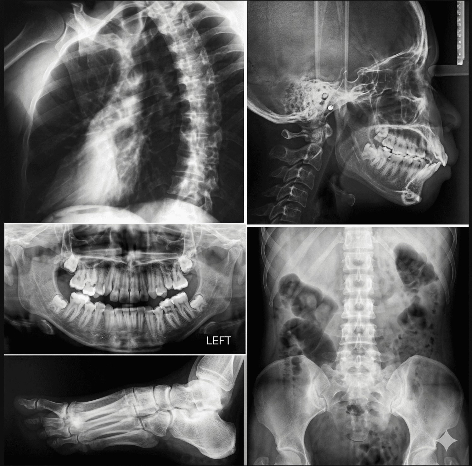
X-Ray
We offer advanced X-ray imaging services, a non-invasive and rapid diagnostic technique that uses low-dose ionizing radiation to visualize internal structures. This modality is highly effective for assessing bones, fractures, infections, and various soft tissue conditions. All examinations are conducted by experienced technologists. The resulting images clearly differentiate structures, with dense tissues such as bones appearing white, and softer tissues appearing in varying shades of grey, enabling accurate and reliable diagnosis.
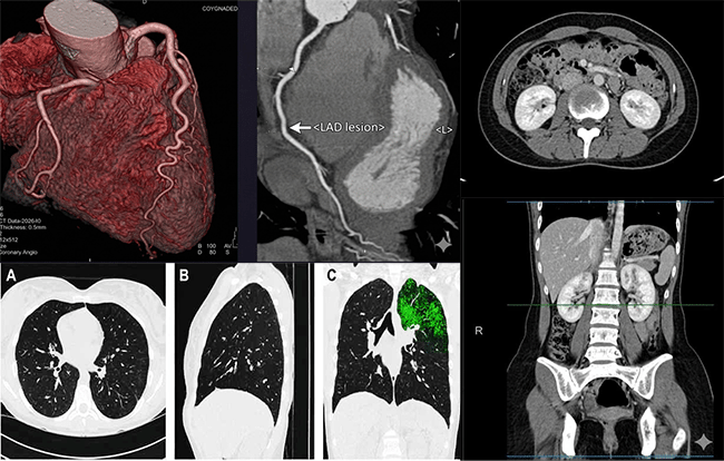
CT Scans
X-MaC provides end-to-end CT scan reporting services for all types of studies, including routine, emergency, and trauma cases, with quick and efficient access. Our subspecialty-trained radiologists ensure fast turnaround times and high-quality, accurate reporting from head to toe, delivering reliable and consistent diagnostic outcomes.
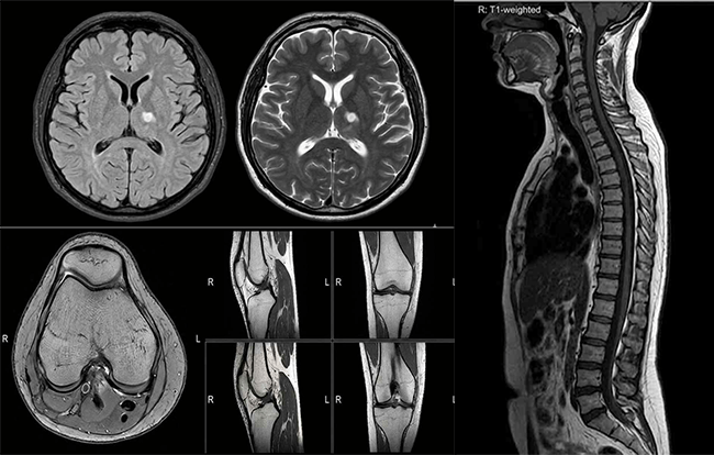
MRI Scans
MRI is a highly specialized area within teleradiology, requiring advanced expertise. At our institution, studies are reported by subspecialty-trained radiologists educated at leading institutes in India and abroad. We offer comprehensive MRI reporting across all major domains, ensuring high standards of accuracy, quality, and clinical reliability.
Cardiac MRI
We provide specialized Cardiac MRI services for detailed, non-invasive evaluation of heart structure, function, and tissue characteristics, aiding in the diagnosis of various cardiac conditions. Our subspecialty-trained radiologists ensure accurate, standardized reporting, delivering reliable insights to support effective cardiac care and treatment planning.
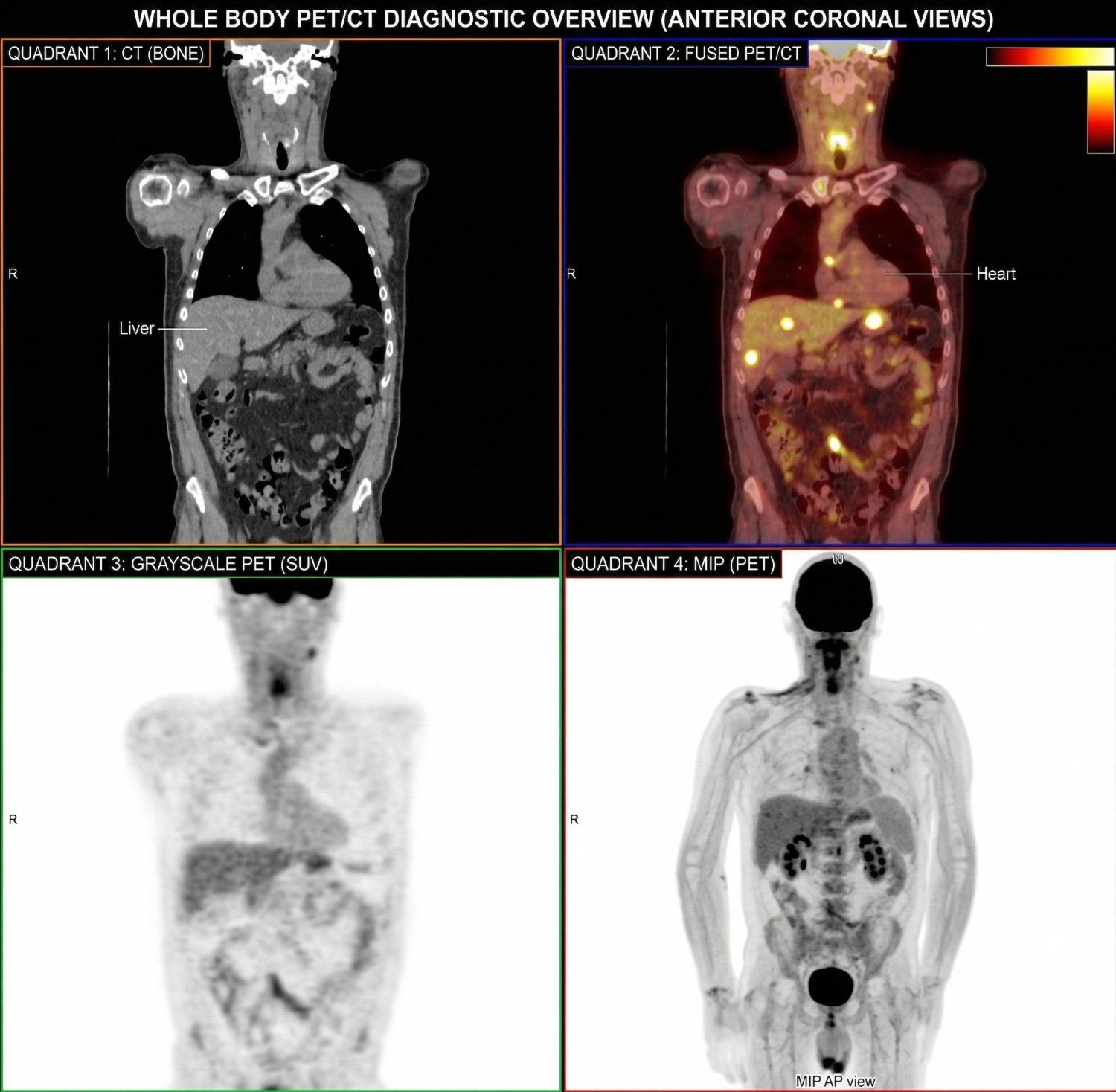
Nuclear Medicine
We also extend our expertise to Nuclear Medicine, offering comprehensive reporting services for all types of gamma scans and PET scans. From oncological imaging to a wide range of functional studies, our experienced Nuclear Medicine physicians ensure accurate and reliable interpretations. Beyond reporting, their specialized expertise supports optimal scan planning, enabling our centers to maximize diagnostic yield and extract clinically meaningful insights from every study.
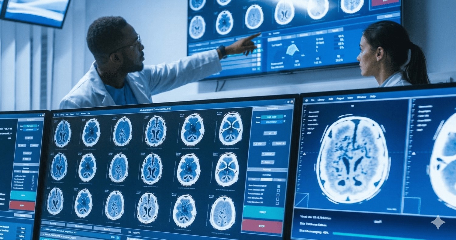
Clinical Discussion
Direct radiologist-to-clinician consultation for complex, ambiguous, or multidisciplinary cases. Real-time telephone and video consultation, MDT meeting participation, and narrative reports with detailed clinical context.
Protocol Optimisation
Evidence-based imaging protocol design and refinement to maximise diagnostic yield, minimise radiation dose, and reduce contrast volume. Audit of existing protocols with improvements aligned to current national and international guidelines.
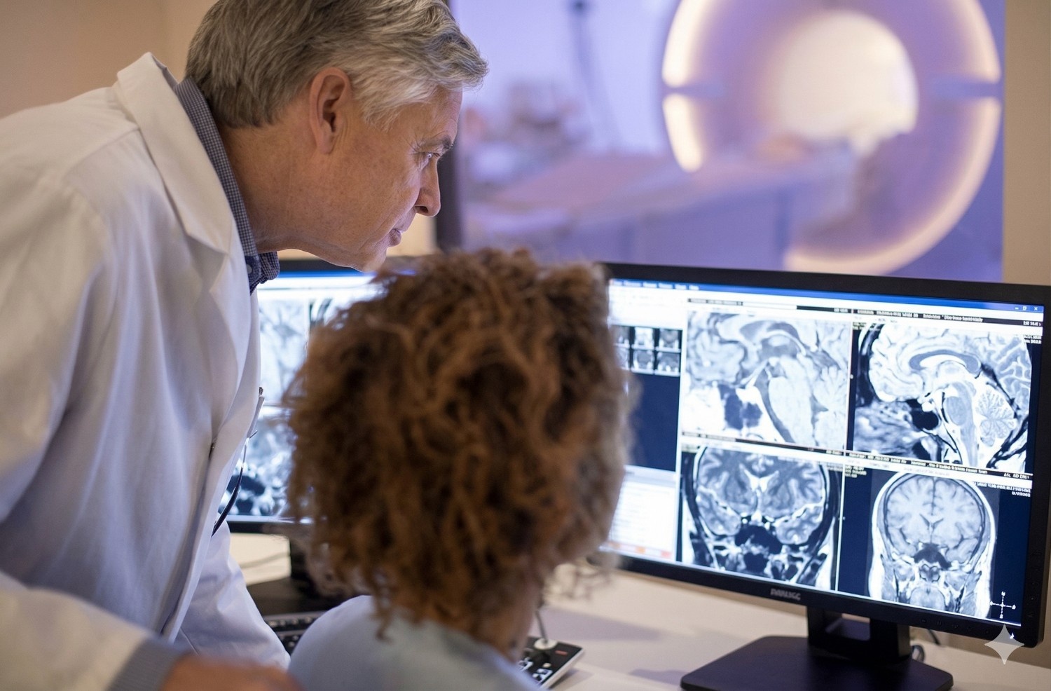
Second Opinion Reporting
We also offer expert second-opinion services for radiology scans, providing critical insights in complex or uncertain cases. Our specialist radiologists conduct a thorough, detail-oriented review to address specific clinical concerns with precision. Additionally, our subspecialty-trained team collaborates closely with referring physicians, discussing findings and contributing to patient management through a multidisciplinary approach.

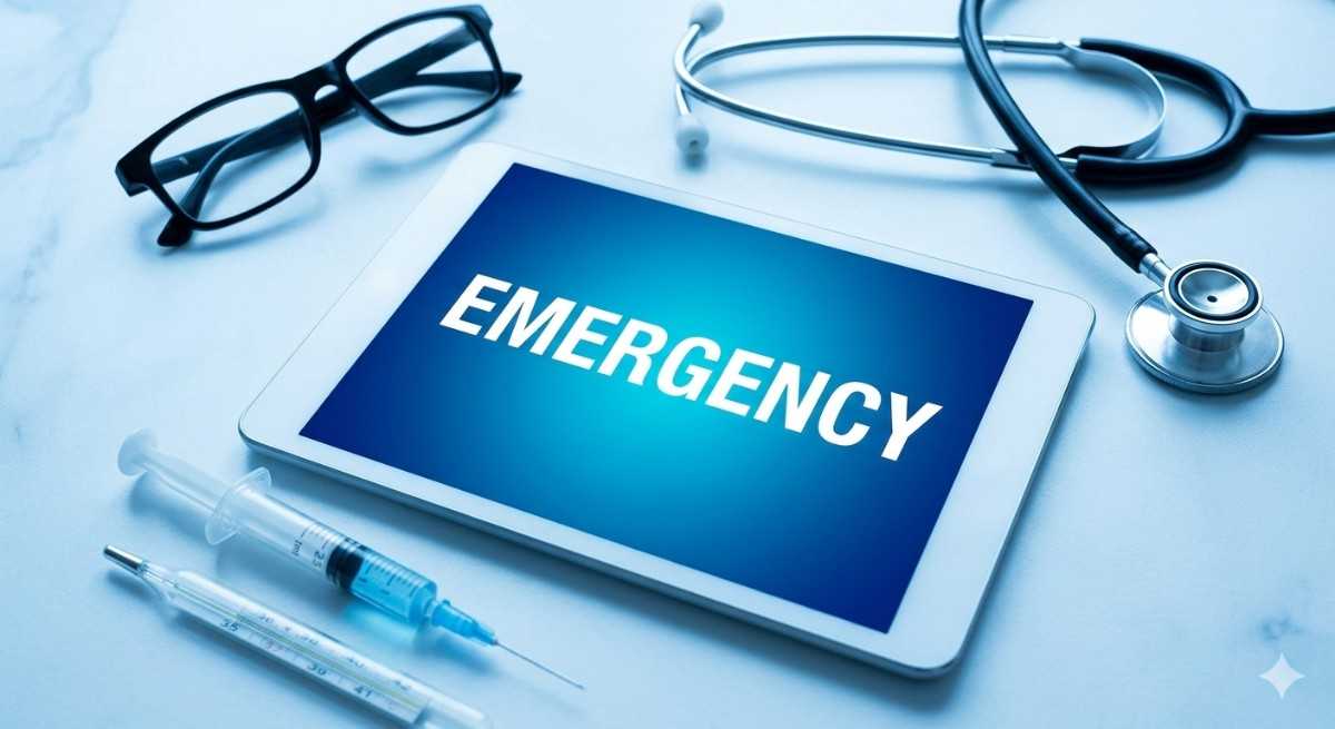
Emergency & Night Reporting
24/7 radiology reporting service for emergency and overnight cases. Fast, accurate reporting for trauma, stroke, ICU, and urgent scans, ensuring continuous support even during night hours. Our expert radiologists deliver timely, high-quality reports with strict turnaround times, helping hospitals maintain uninterrupted workflow. Seamless integration, data security, and reliable communication ensure confidence in every critical decision.